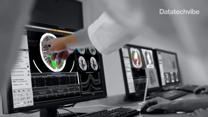
Offering more than 70 clinical applications, the next-generation edition of the Advanced Visualisation Workspace reportedly includes enhanced liver analysis and AI-powered scoring of early brain infarction noted on computed tomography (CT) scans for patients with ischemic stroke.
Emphasising a variety of AI algorithms and workflows across multiple imaging modalities and disciplines on one platform, Philips launched a new edition of the Advanced Visualisation Workspace at the Radiological Society of North America (RSNA) annual conference in Chicago.
With the goals of streamlined radiology workflows and improved diagnostic confidence, the Advanced Visualisation Workspace provides a suite of enhanced imaging options with more than 70 clinical applications for radiology, oncology, neurology, and cardiology, according to the company.
Philips said the new applications include:
- CT ASPECT (Alberta Stroke Program Early CT Score), an AI-powered tool that enables the scoring of early brain infarction on non-contrast CT exams for patients who have had an ischemic stroke.
- MR cardiac suite that provides diagnostic support and facilitates reporting with one overview of all imaging data types.
- CT Liver Analysis allows analysis and quantification of liver segments and other defined regions of interest.
“At this year’s RSNA, Philips will showcase how our informatics solutions use intelligence to provide patient-centric insights, integrate advanced visualisation tools into the workflow and support clinical collaboration to speed up the detection of diseases by leveraging intelligence everywhere along the patient care journey,” noted Reema Poddar, the president of Diagnostic and Pathway Informatics at Philips.


